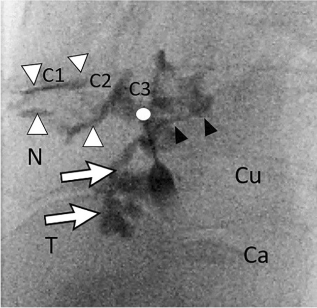
Fig. 27.

A 75-year-old female with history of multiple sprains and midfoot osteoarthritis, referred for naviculocuneiform (NC) CSI. Fluoroscopic image shows the needle targeting the lateral NC joint (circle), due to extensive osteophyte formation medially. As expected, contrast disperses into all three NC articulations and recesses (arrowheads). In addition, there is abnormal, unexpected contrast dispersal proximally into the talonavicular joint recess (arrows), and into the cuboid-lateral cuneiform joint (black arrows) due to prior capsular injury. In this case, the collimated image does not include the TMT joints, where we would expect to see normal extension of contrast into the 2nd and 3rd TMT joints. Lobulated filling defects imply synovitis. T = talus. N = navicular. Ca = calcaneus. Cu = cuboid. C1–C3 = medial, middle, and lateral cuneiforms, respectively
