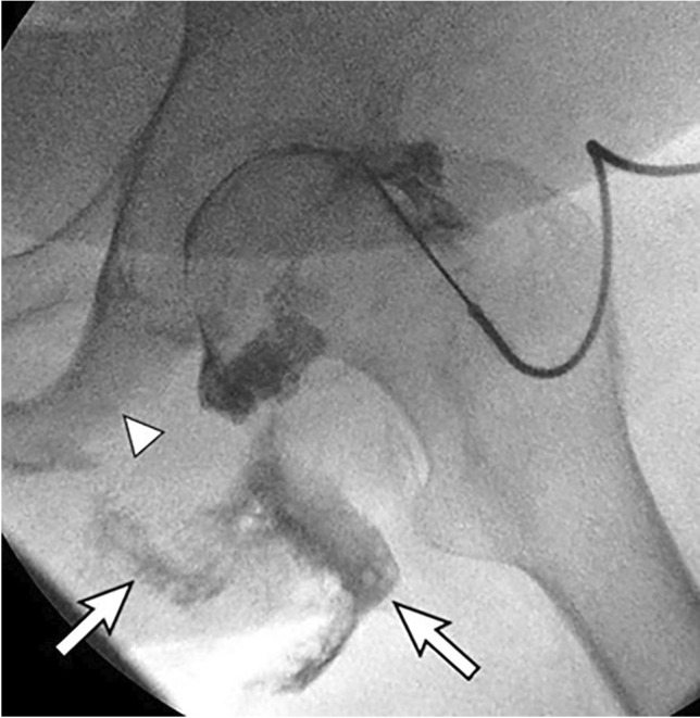
Fig. 5.

A 69-year-old male with left ischial decubitus ulcer with suspected septic left hip joint. Aspiration attempts yielded no fluid. Fluoroscopic image shows contrast injection not only opacifying the left hip joint but also extending into the ischial decubitus ulcer (arrows). The ischial tuberosity is eroded (arrowhead), consistent with osteomyelitis
