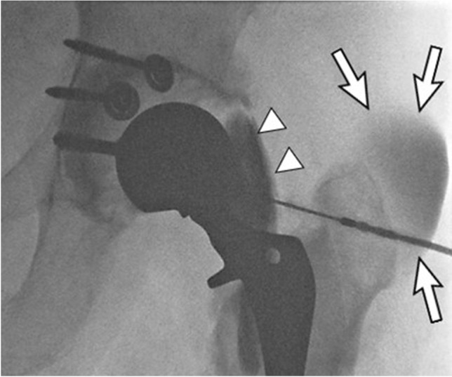
Fig. 9.

A 60-year-old male with infected left total hip arthroplasty, explanted acetabular component with acetabular spacer placement. Follow-up aspiration yielded a dry tap. Fluoroscopic image of the left hip after subsequent contrast injection shows communication of the hip joint with the iliopsoas bursa (arrowheads) and contrast decompressing into an abscess overlying the greater trochanter (arrows)
