Figure 3.
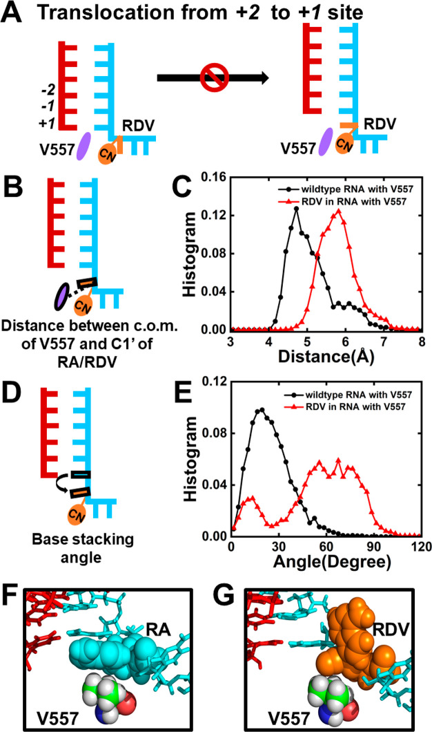
Translocation of RDV from the +2 to +1 site is hindered by V557. (A) Diagram showing V557 hampers the translocation of RDV from the +2 to the +1 site, where RDV and V557 are colored in orange and purple, respectively. (B) Cartoon model showing the distance between the center of mass (c.o.m.) of V557 and C1′ of RA/RDV. (C) Histogram of the distance as shown in (B) when RA (black) or RDV (red) translocates. (D) Cartoon model showing the base-stacking angle between RDV/RA and the upstream nucleotide. (E) Histogram of the base-stacking angle for RA (black) or RDV (red) during translocation. (F,G) Typical conformation of RA/RDV with V557 during translocation.

