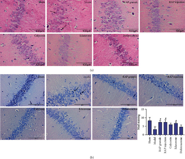
Figure 4.

(a) Representative images of HE staining of the hippocampal CA3 area of the ischemic side in the rats (×400). (b) Representative images of Nissl staining in the hippocampal CA3 region (×400). Black arrow: necrotic neuron. Scale bar = 100 μm. Statistical results of the Nissl staining in the seven groups (n = 5). ∗∗P < 0.01 vs. sham group, #P < 0.05 vs. model group.
