
Figure 1.
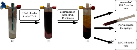
PRP preparation process by Mini GPS III. (a) The empty Mini GPS III tube. (b) The tube filled with 3 ml of anticoagulant citrate dextrose solution A (ACD-A) and 27 ml of patients' own blood. (c) The tube after centrifugation containing three separate layers: platelet-poor plasma (PPP), platelet-rich plasma (PRP), and red blood cells (RBC). (d) The syringe filled with PRP taken from the tube after the removal of PPP.