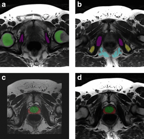
Fig. 1.

a, b Two slices of the transversal multi-echo spin echo (MESE) image (TE = 106 ms) of an asymptomatic volunteer, with manual delineations within the reference regions. Purple indicates the obturator internus muscle, yellow the ischial tuberosity (pelvic bone), blue the ischioanal fossa (fat) and green the yellow bone marrow in the femoral heads. c Transversal T2-weighted image registered to the MESE image space, with co-registered prostate segmentation. The peripheral zone is red, while the remaining zones (transitional zone, central zone and anterior fibromuscular stroma) are green. d Transversal MESE image (TE = 106 ms) with registered manual prostate segmentations
