Figure 7.
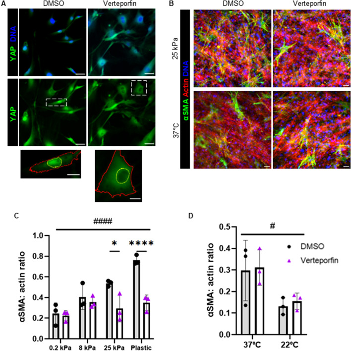
Yap inhibition reduces myofibroblast activation. (A) YAP localization in FAPs cultured on plastic in DMSO or 0.5 µM verteporfin for 2 days. Scale bars are 50 µm, insets are 20 µm. Yellow outlines indicate nuclei and red outlines indicate cytoplasm on insets. (B) Myofibroblast activation in FAPs cultured for 7 days with 0.5 µM verteporfin or 0.1% DMSO on collagen-coated polyacrylamide gels at 25 kPa and 3.0 mg/ml telocollagen gels polymerized at 37 °C. Scale bars set to 50 µm. (C) Quantification of myofibroblast activation of FAPs in DMSO or 0.5 µM verteporfin on collagen-coated polyacrylamide gels. (D) Quantification of myofibroblast activation of FAPs in DMSO or 0.5 µM verteporfin on collagen gels polymerized at different temperatures. *P < 0.05, ****P < 0.0001 between DMSO and verteporfin treatment. #P < 0.05, ####P < 0.0001 between substrates.

