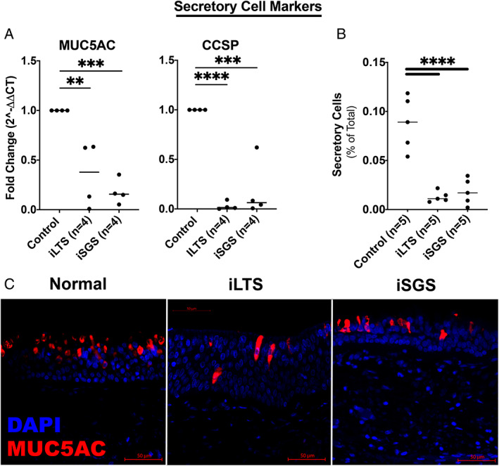
Fig. 6.

Low secretory cell expression in iSGS and iLTS‐scar—(A) MUC5AC and CCSP, two secretory cell markers were significantly reduced in both iSGS and iLTS‐scar epithelium compared with non‐scar controls. (B) Secretory cells counted on H&E sections were significant reduced in both iSGS and iLTS‐scar epithelium. (C) Representative immunohistochemical staining of MUC5AC demonstrated reductions in goblet cells and changes in morphology in iLTS and iSGS‐scar epithelium. * = P < .05; ** = P < .01; *** = P < .001; **** = P < .0001. [Color figure can be viewed in the online issue, which is available at www.laryngoscope.com.]
