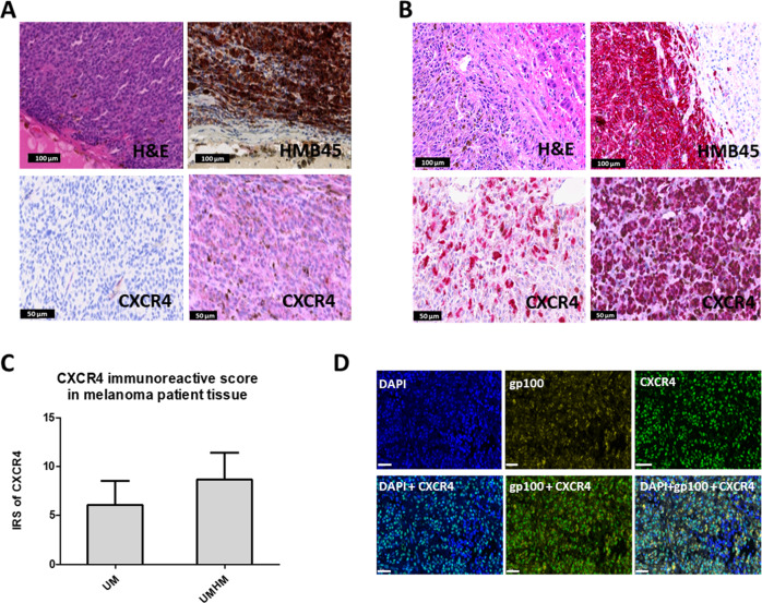
Fig. 1. Evaluation of CXCR4 expression levels in primary UM and hepatic metastases of UM (HMUM).
A Histological analysis of primary UM tissue. H&E staining (top left) shows the morphology of UM, and the diagnosis of UM is confirmed by melanoma marker Human Melanoma Black (HMB45, brown) IHC staining (top right). CXCR4 IHC staining shows low (bottom left) to moderate (bottom right) expression level in UM tissues. B Histological analysis of HMUM. H&E (top left) and HMB45 (top right) IHC staining (red) confirmed the lesion as UM metastases in the liver. CXCR4 IHC staining (bottom left and bottom right) shows high expression levels (denoted by red staining) in HMUM. C Immunoreactive score (IRS) of CXCR4 in primary UM (UM) and hepatic metastasis of UM (HMUM). HMUM has significantly higher CXCR4 IRS than UM (P < 0.01). D Immunofluorescence staining of DAPI (blue, top left), gp100 (yellow, top center), and CXCR4 (green, top right) in UM hepatic metastases tissue (scale bars, 60 µm). The spatial overlapping of gp100 and CXCR4 (bottom center) demonstrated CXCR4 expressed on tumor cells (scale bars, 60 µm). Bottom left: the merged image of CXCR4 and DAPI; bottom right: the merged image of CXCR4, gp100 and DAPI.

