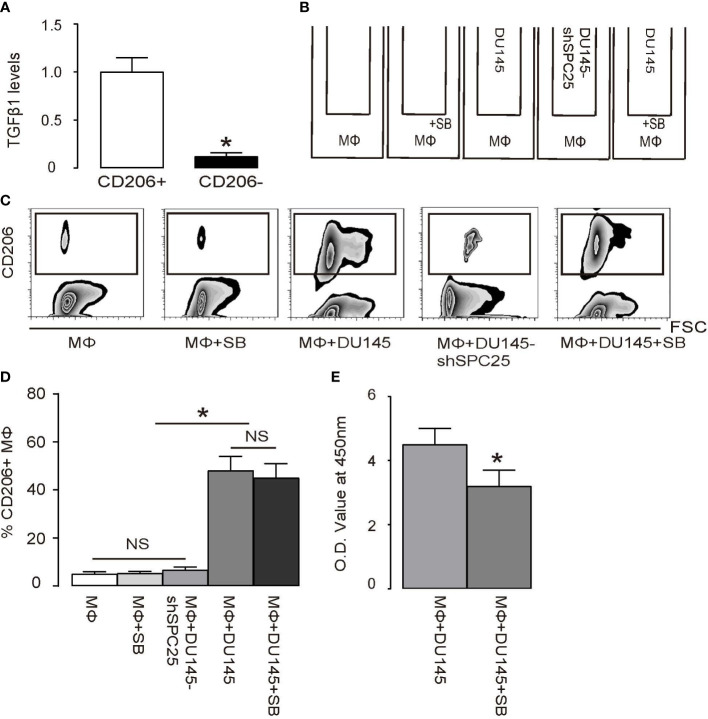
Figure 5.
Polarized TAMs promote PrC cell growth through TGFβ1 (A) RT-qPCR for TGFβ1 mRNA level in CD206+ versus CD206- macrophages. (B) A transwell co-culture diagram of DU145 cells and bone-marrow-derived macrophages (MΦ). Group 1: MΦ. Group 2: MΦ with inhibitor SB431542 (SB). Group 3: MΦ and DU145 cells. Group 4: MΦ and DU145 cells transduced with shSPC25. Group 5: MΦ and DU145 cells with SB. Cells were incubated for 2 days. (C, D) Flow cytometry analysis of CD206+ cells in macrophages, shown by quantification (C) and by representative flowcharts (D). (E) MTT assay on DU145 cells. *p < 0.05. NS, non-significant. n = 5.

