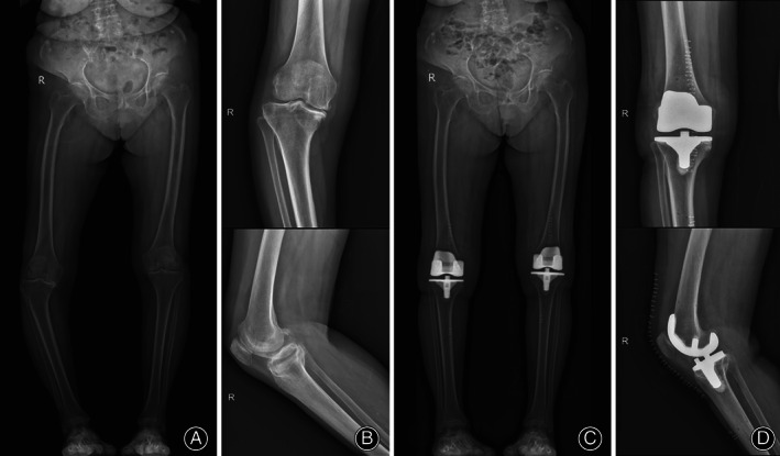
Fig. 1.

Radiographs of an 80‐year‐old female patient who underwent navigation‐assisted TKA. The arKA technique was performed on the right knee and aMA was used on the left knee. (A) Preoperative standing full‐lower limb X ray (EOS) showed an osteoarthritis right knee with 153.65° severe varus deformity. (B) Preoperative X‐ray of the right severe varus knee. (C) Postoperative EOS showed a restored right knee with an angle of 5.87° varus and a coronal femoral component angle (β) of 91.29° with a coronal tibial component angle (γ) of 86.98°. (D) Postoperative X‐ray showed a sagittal femoral component angle (δ) of 0.71°, tibial posterior slope angle (ε) of 89.47°, and femoral‐patella angle (θ) of 30.57°
