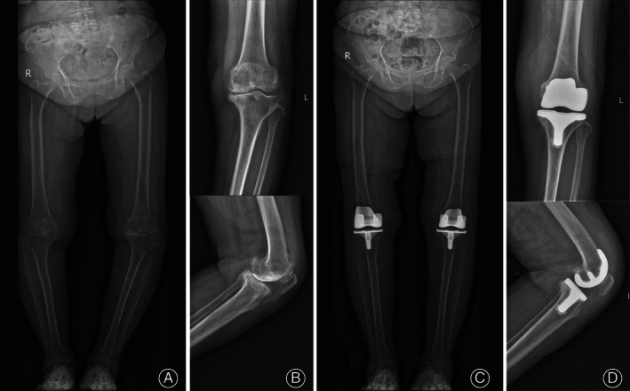
Fig. 3.

Radiographs of a 73‐year‐old female patient who underwent TKA using arKA on the left knee and rKA on the right knee. (A) Preoperative standing full‐lower limb X ray (EOS) showed a severe varus deformity of 156.10° on the left knee. (B) Preoperative X‐ray of the left severe varus knee. (C) Postoperative EOS showed a restored left knee with an angle of 6.17° varus and a coronal femoral component angle (β) of 93.30° with a coronal tibial component angle (γ) of 86.83°. (D) Postoperative X‐ray showed a sagittal femoral component angle (δ) of 1.00°, tibial posterior slope angle (ε) of 88.76°, and femoral‐patella angle (θ) of 30.86°
