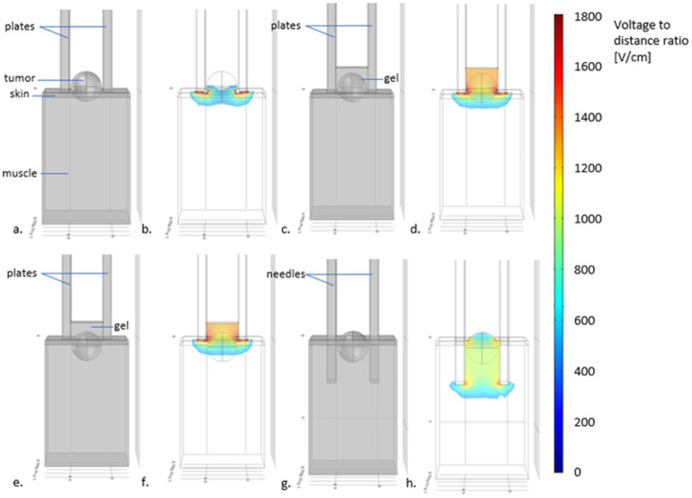
Figure 5.
Scheme displaying the depth of treatment with different electrodes and visual recommendations for the use of plate or needles electrodes. In (a) plates electrodes used for treating a superficial tumor, without using conductive gel. In (b) the simulation of the electric field distribution (using COMSOL Multiphysics 4.3—in color the electric field intensities above the threshold for tissue reversible electroporation) reveals that the superficial parts of the tumor may not be adequately treated. In (c) the scheme shows the addition of gel between the plates. In (d), the simulation shows that now, the distribution of the electric field allows treating the tumor completely. In (e), the case of a tumor with an invasion depth >3 mm treated with non-penetrating plate electrodes. As it can be seen in (f), even with the use of gel, the deepest parts of the tumor may not be adequately treated. Indeed, the field intensity drops below the electroporation threshold at a distance lesser than the separation of the plates. For these cases, needle electrodes should be used, as depicted in (g) where the same tumor (with an invasion depth >3 mm) is treated with needle electrodes without conductive gel. The electric field simulation in (h) shows that the whole tumor is now completely treated.

