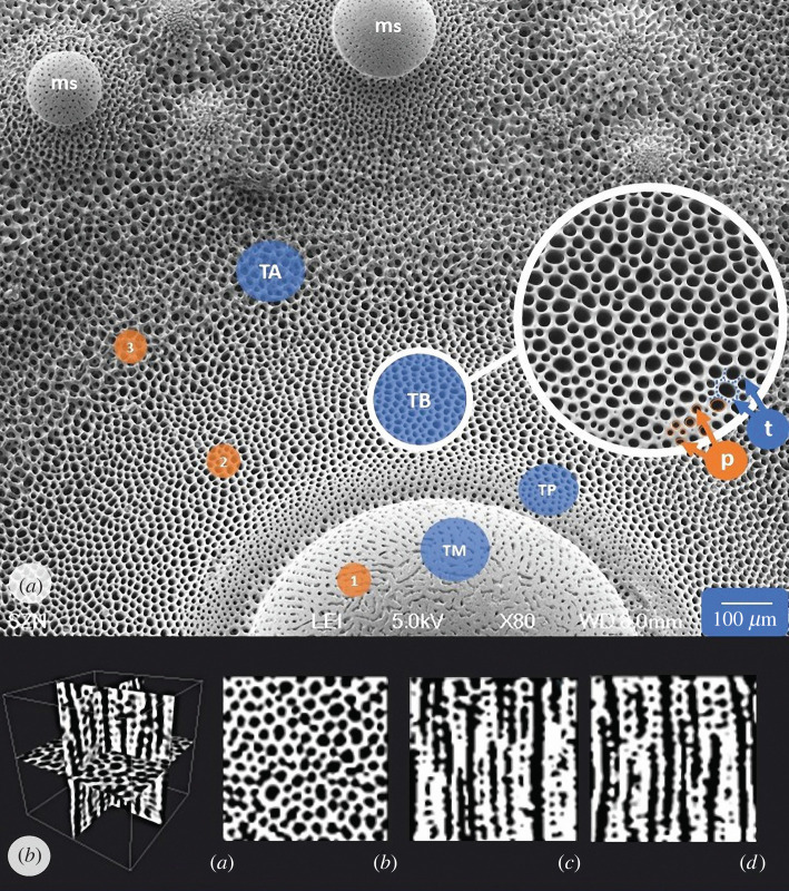
Figure 2.
Tubercle architecture and regions examined. (a) SEM micrograph (top view) of Paracentrotus lividus interambulacral plate showing the primary spine tubercle and its stereom microstructural variability. Three stereom types can be recognized: (1) microperforate, (2) galleried, and (3) labyrinthic. In addition, the mamelon of secondary spines (ms) is also shown. The region topographic reference is underlined by a solid line circle in which the pores (p) and trabeculae (t) are indicated (arrows). (b) Micro-CT scan of tubercle boss subsection extracted by P. lividus interambulacral plate showing (a) transversal, (b) sagittal and (c) coronal views.

