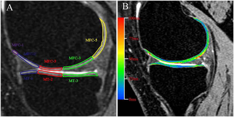
Figure 4.
(A) Regions of interest (ROI) segmentation of the medial tibiofemoral compartment. The MFC and MT were segmented manually into 5 and 3 subregions, respectively, according to the meniscus. Each subregion was assessed by further segmenting full-thickness cartilage into 2 approximately equal sections. MFC-3 and MT-2 are contacting regions of femoral and tibial cartilage during standing. MFC-2, MFC-4 and MT-1, MT-3 are regions above and below the meniscal horn, respectively. MFC-1 and MFC-5 are non-weightbearing portions of the femoral condyle during standing. T2 mappings of the MMPRTs after surgery. (B) Mean T2 values from superficial and deep layers in each defined subregions, as shown in (A), were recorded. MFC = medial femoral condyle; MT = medial tibia; MMPRTs = medial meniscus posterior root tears.

