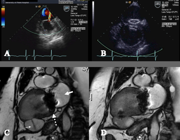
Figure 4.
Percutaneous repair of left ventricular pseudoaneurysm. A. Transesophageal echocardiogram with color Doppler flow showing a thinned inferior wall infarction with a jet of flow into the large pseudoaneurysm; B. Transesophageal echocardiography showed the two discs of the occluder device seated across the defect. Spontaneous echo contrast (“smoke”) indicated stasis in the pseudoaneurysm; C. Cardiac MRI showing akinetic inferior wall segment with a jet of flow into the pseudoaneurysm. The bioprosthetic mitral valve is seen. (Bold white arrow = Left atrium; Narrow white arrow = pseudoaneurysm; dotted white arrow = mitral valve.); D. Cardiac MRI seven days after implantation of the occluder device showed no flow into the pseudoaneurysm.

