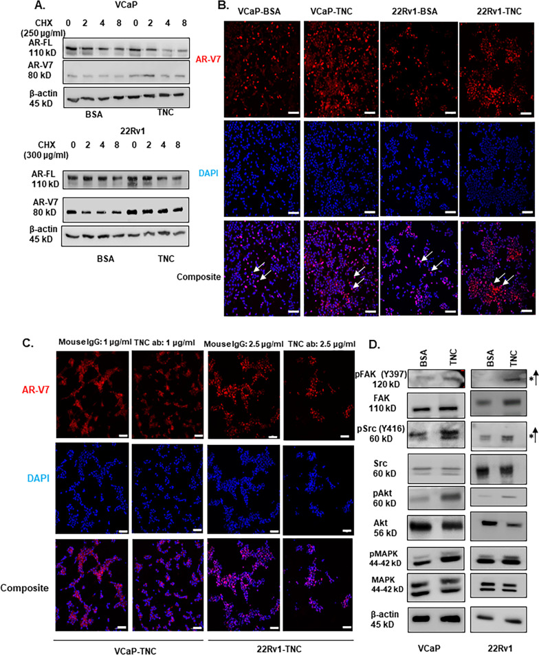
Fig.4.
TNC induce post-translational stability of AR-V7 and activate FAK and Src signaling. A VCaP and 22Rv1 cultured on BSA versus TNC in 5% csFBS containing media were treated with cycloheximide (VCaP-250 μg/ml; 22Rv1-300 μg/ml) and Western blot conducted on protein lysate collected at 0,2,4, and 8 h. The cycloheximide chase experiment was conducted for n = 3 biological replicates for each cell line. B ICC of AR-V7 nuclear localization (white arrow) in both VCaP and 22Rv1 cultured on TNC compared to BSA coated IbiTreat chamber slides (Scale bar, 10 × 100 µm). C VCaP and 22Rv1 were cultured on TNC coated IbiTreat chamber slides followed by treatment with isotype control (IgG) or anti-tenascin monoclonal antibody (BC-24) at a concentration of 1 µg/ml (VCaP) and 2.5 µg/ml (22Rv1) respectively for 72 h. The nuclei are counterstained with DAPI. All ICC images represented in Fig. 4. were obtained using Nikon A1 confocal microscope (Scale bar, 10 × 100 µm) and repeated for n = 3 biological replicates. D Western blot analysis FAK, Src, Akt, and MAPK phosphorylation status in VCaP and 22Rv1 cultured on BSA versus TNC for n = 3 biological replicates

