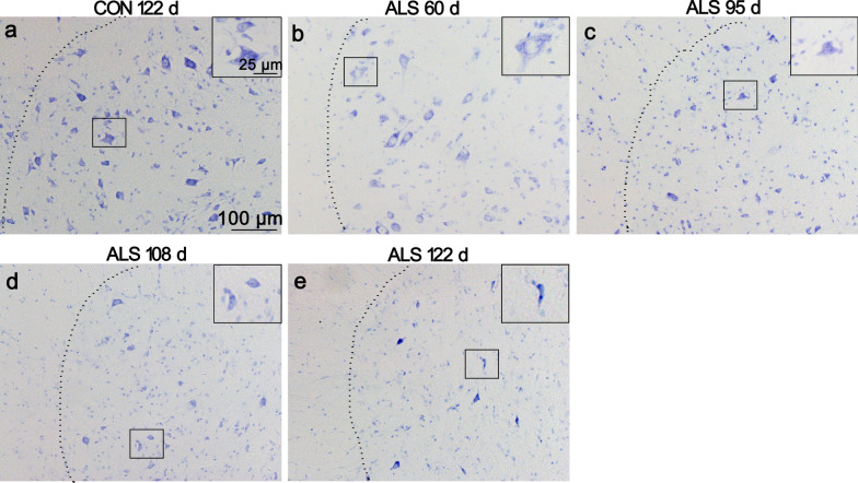
Fig. 1.
Neural degeneration in the ventral horn of ALS mice lumbar spinal cord. Representative photomicrographs of Nissl-stained lumbar sections of 122 d CON mice a, 60 d ALS mice b, 95 d ALS mice c, 108 d ALS mice d and 122 d ALS mice (e, scale bar 100 μm) are shown. Inserts depict higher magnifications of the overview images (Scale bar 25 μm). In ALS mice, altered neurons were frequently observed from 60 d of age with vacuolation in the soma. With disease progression, spongiform degeneration and loss of large motoneurons was detected.

