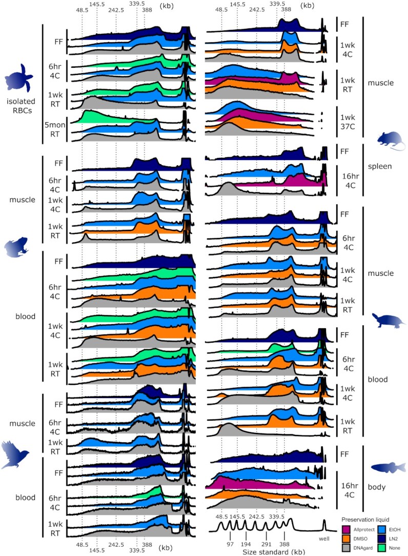
Figure 2:

Pulsed-field gel electrophoresis (PFGE) measurements of uHMW DNA comparing different sample temperature and storage times. PFGE traces are visualized as overlapping ridgeline plots. Fluorescent-stained DNA fragments are drawn with an electric current from the well at the right toward the left. Smaller fragments generally travel farther than larger fragments. The fragments that greatly exceed the targeted size range remain in the well and cannot be reliably interpreted. Each ridgeline plot corresponds to a gel lane and a single DNA extract with brightness converted to a plot profile. The x-axis denotes molecule length scaled via piecewise linear scaling to match across gels of different lengths with a common size standard (Lambda PFG Ladder, New England Biolabs). The x-axis is the same in both columns. The y-axis of each plot is brightness scaled proportionally in each gel lane from just below the well to just beyond the 48.5-kb ladder peak such that the relatively intense brightness of the well itself is excluded from scaling. The well brightness is cropped where it exceeds the brightness of the rest of the gel lane. DNA fragments with lengths longer or shorter than peaks of the size standard cannot be reliably interpreted due to lack of size reference and artifacts of gel electrophoresis as well as limitations of any type of gel electrophoresis to correctly size megabase-length fragments. Colors represent different sample preservation methods, as indicated in the legend at bottom right. All samples were transferred to –80°C after the specified temperature treatment (e.g., “6hr 4C” means stored at 4°C for 6 hours before transfer to –80°C). Abbreviations are as follows: RBCs, isolated red blood cells; EtOH, 95% ethanol; DMSO, a mix of 20–25% dimethyl sulfoxide, 25% 0.5 M EDTA, and 50–55% H2O, saturated with NaCl. DNAgard, DNAgard tissue and cells (cat. no. #62001–046; Biomatrica); Allprotect, Allprotect Tissue Reagent (cat. no. 76405; Qiagen); FF, flash-frozen in liquid nitrogen immediately upon dissection; 6hr, 6 hours; 1d, 1 day; 1wk, 1 week; 5mon, 5 months; RT, room temperature (20–25°C). Three additional samples were tested but produced insufficient DNA for fragment length analysis: frog muscle in DMSO for 1 week at 4°C and 6 hours at 4°C and mouse spleen in DMSO for 16 hours at 4°C. For measurements based on the FEMTO pulse instrument and additional tissue types, see Supplementary Figs. S2 and S3.
