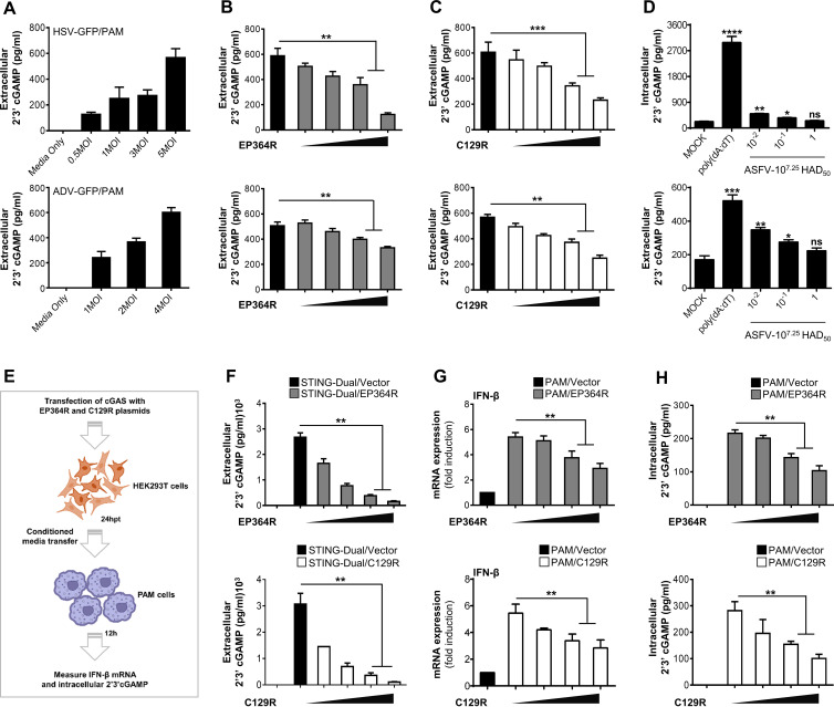
FIG 4.
EP364R and C129R degrade 2′,3′-cGAMP and impair its transfer to bystander cells. (A) PAMs were infected with HSV-GFP (MOI = 0.5, 1, 3, and 5) and ADV-GFP (MOI = 1, 2, and 4) for 4 h and 2′,3′-cGAMP level in the supernatant was analyzed using ELISA. (B and C) PAMs were cotransfected with Flag-EP364R (B) or Flag-C129R (C) plasmids dose dependently. Cells were then infected with HSV-GFP (MOI = 5) and ADV-GFP (MOI = 4) for 4 h and assessed the 2′,3′-cGAMP secretion by ELISA. (D) Primary porcine alveolar macrophages were either mock infected or infected with 10-fold-diluted 107.25 HAD50 of ASFV-WT as indicated. Poly(dA-dT) was transfected as a positive control. At 5 hpi or 5 hpt, intracellular and extracellular cGAMP levels were measured. (E) Graphical illustration of conditioned-medium transfer experiment. (F) 293-Dual hSTING-A162 cells were cotransfected with Flag-EP364R or Flag-C129R dose dependently with 3×Flag-cGAS plasmid with Flag vector as a transfection control, and the 2′,3′-cGAMP secretion was determined at 24 hpt in the supernatant by ELISA. (G and H) Another 5(F)-independent experiment cell supernatant was collected and immediately transferred to PAMs. After 24 h of incubation, IFN-β transcription (G) and intracellular 2′,3′-cGAMP level (H) were measured compared to untreated control cells. In panel G, data are representative of at least two independent experiments. ELISA and GFP absorbance data are representative of at least two independent experiments, each with similar results, and the values are expressed as means and SD for three biological replicates. Student's t test: **, P < 0.01; ***, P < 0.001; ****, P < 0.0001.

