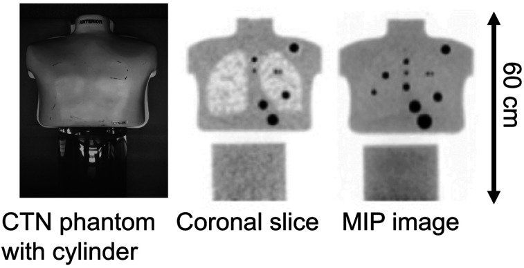
FIGURE 1.
Photograph of CTN torso phantom and 20-cm-diameter by 30-cm-long cylinder used for these measurements (left), along with example reconstructed coronal (middle) and maximum-intensity-projection (MIP) (right) images. Lip at bottom of phantom and top of cylinder shows as gap in reconstructed images. Spheres in these images are physical spheres that are part of phantom and were not used for detectability study.

