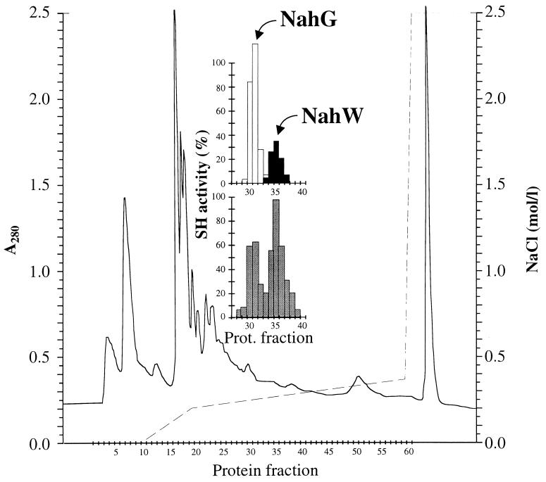
FIG. 5.
Anion-exchange chromatography of extracts of salicylate-induced P. stutzeri AN10 cells. Protein was monitored as A280 (solid line). Gradient elution with NaCl (broken line) was used. (Inset) Salicylate hydroxylase activity was determined as NADH oxidation, as described in Materials and Methods, using salicylate (grey bars), 3-methylsalicylate (white bars), and 3-chlorosalicylate (black bars) as substrates.

