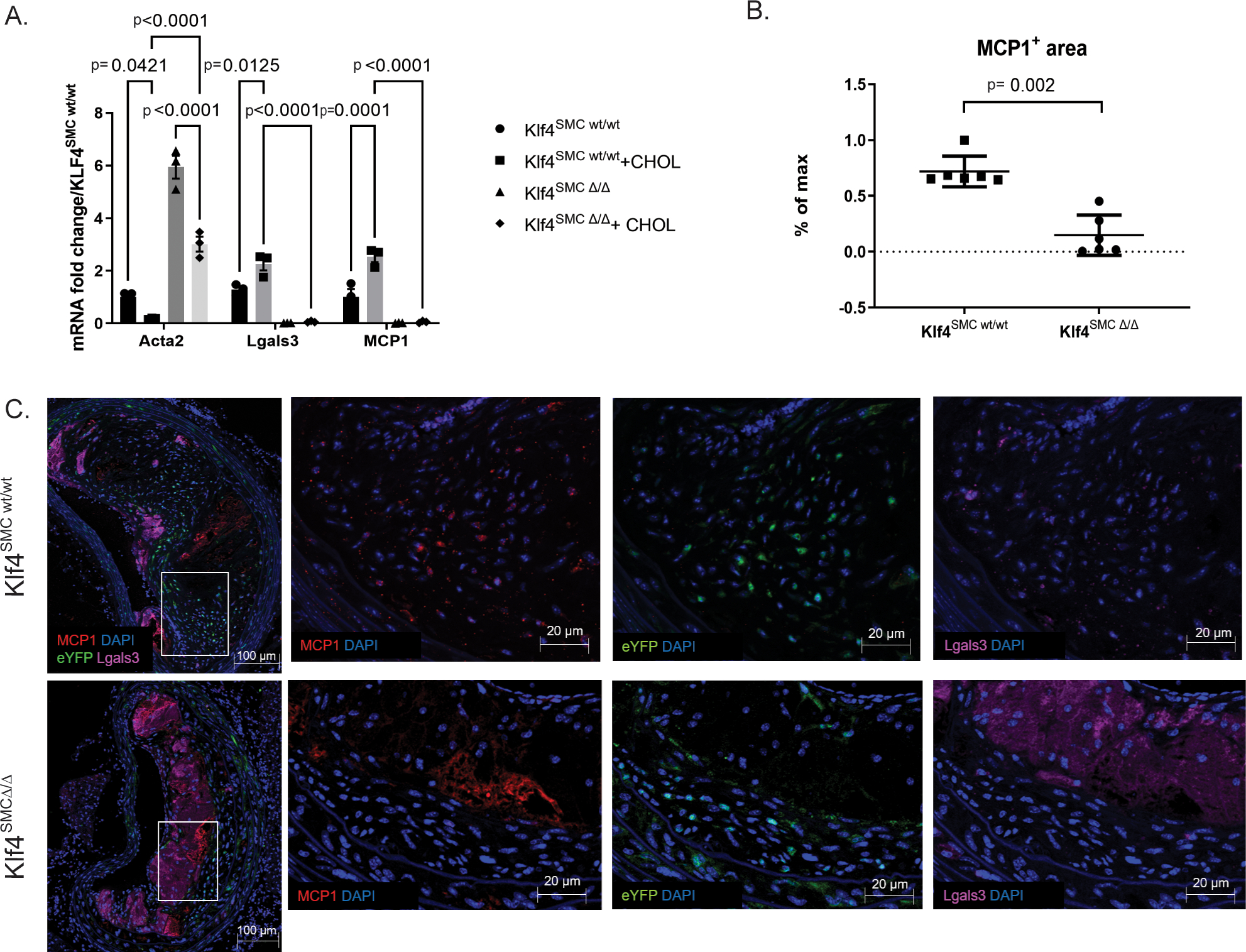
Figure 1. Smooth muscle cells express MCP1 in an Klf4-dependent manner.

A) Cultured aortic SMC from SMC lineage-tracing mice on an ApoE background with or without SMC specific Klf4 knockout (Klf4SMC) were were treated with 0.2% FBS +/− 80 ng/μL cholesterol media for three days, and qRT-PCR was performed for SMC and macrophage marker genes. n=3, comparisons by two-way ANOVA and Tukey’s multiple comparisons test. B) After 18 weeks of Western diet feeding, brachiocephalic artery (BCA) lesions collected from Klf4SMC wt/wt and Klf4SMC Δ/Δ mice were stained for eYFP(marking lineage-traced SMC), LGALS3, MCP1, and DAPI. MCP1+ area per lesion quantified by pixilation. n=6 mean +/− SEM. p<0.001 by Mann Whitney test C) Representative images are shown from the experiment in B.
