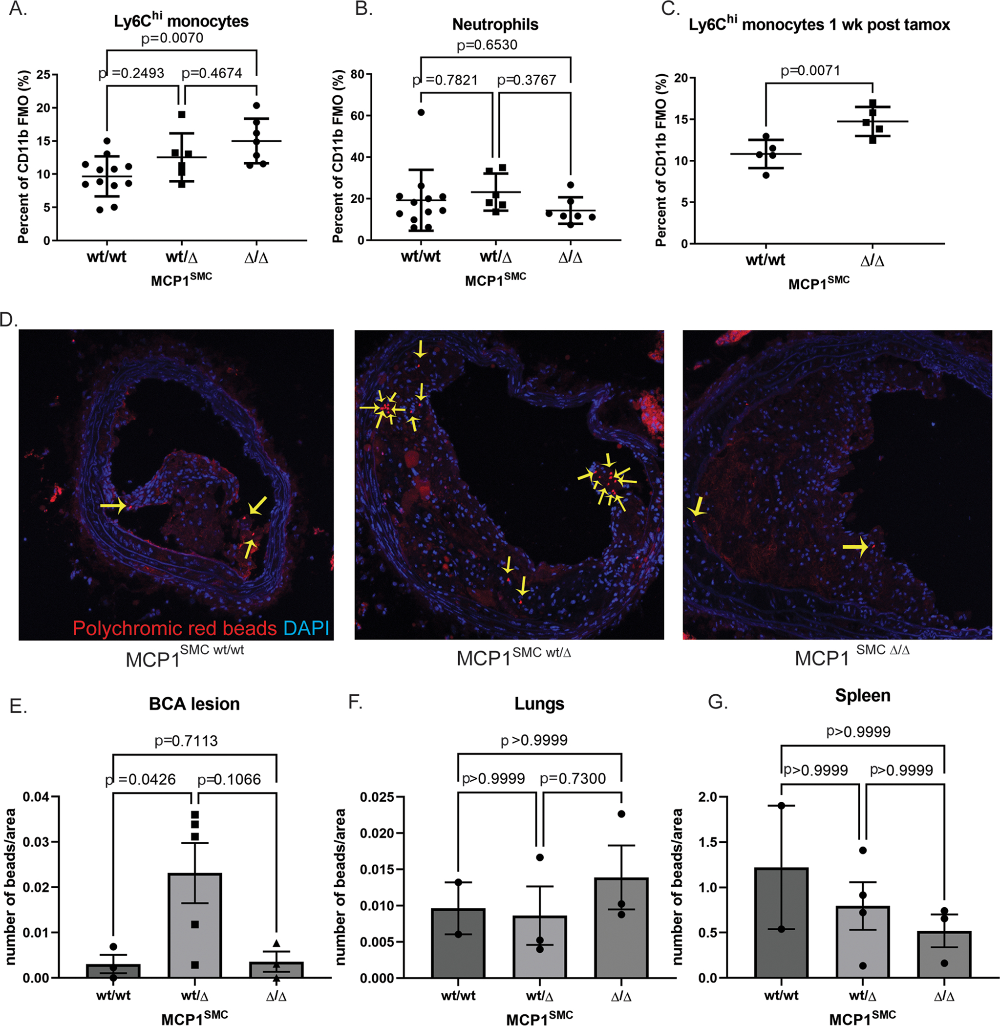
Figure 3. MCP1SMC wt/Δ and MCP1SMC Δ/Δ mice have significantly more Ly6Chi monocytes in peripheral blood.

A-C Peripheral blood from MCP1SMC wt/wt, MCP1SMC wt/Δ and MCP1SMC Δ/Δ mice was collected after 18 weeks of Western diet (A-B) or by retro-orbital bleed 1 week after tamoxifen injection (C), stained for leukocyte markers, and analyzed by flow cytometry. Cells were gated on live, single CD45+CD11b+ Ly6G- (A,C) or Ly6G+ cells (B) with fluorescence-minus-one controls. n=5–9, mean +/− SEM. P values represent pairwise Sidak’s multiple comparison test after one way ANOVA for A and B, and C is a Mann-Whitney test. D) MCP1SMC mice fed 15 wks Western diet were tail-vein injected with fluorescent beads to label circulating monocytes. BCA plaques were collected 3 weeks later after 18 weeks of Western diet and frozen sections were imaged for beads (yellow arrows) within lesion cells. Representative images from this experiment are shown in D. E) Beads from the experiment in D were quantified as a percent of lesion area and analyzed by Kruskal-Wallace test with multiple comparisons. F-G) Bead content per area was also measured in non-atherosclerotic lung tissue to assess possible changes in overall tissue monocyte flux and in spleens to measure monocyte efflux. Three locations 100 μm apart were sampled and averaged for each animal.
