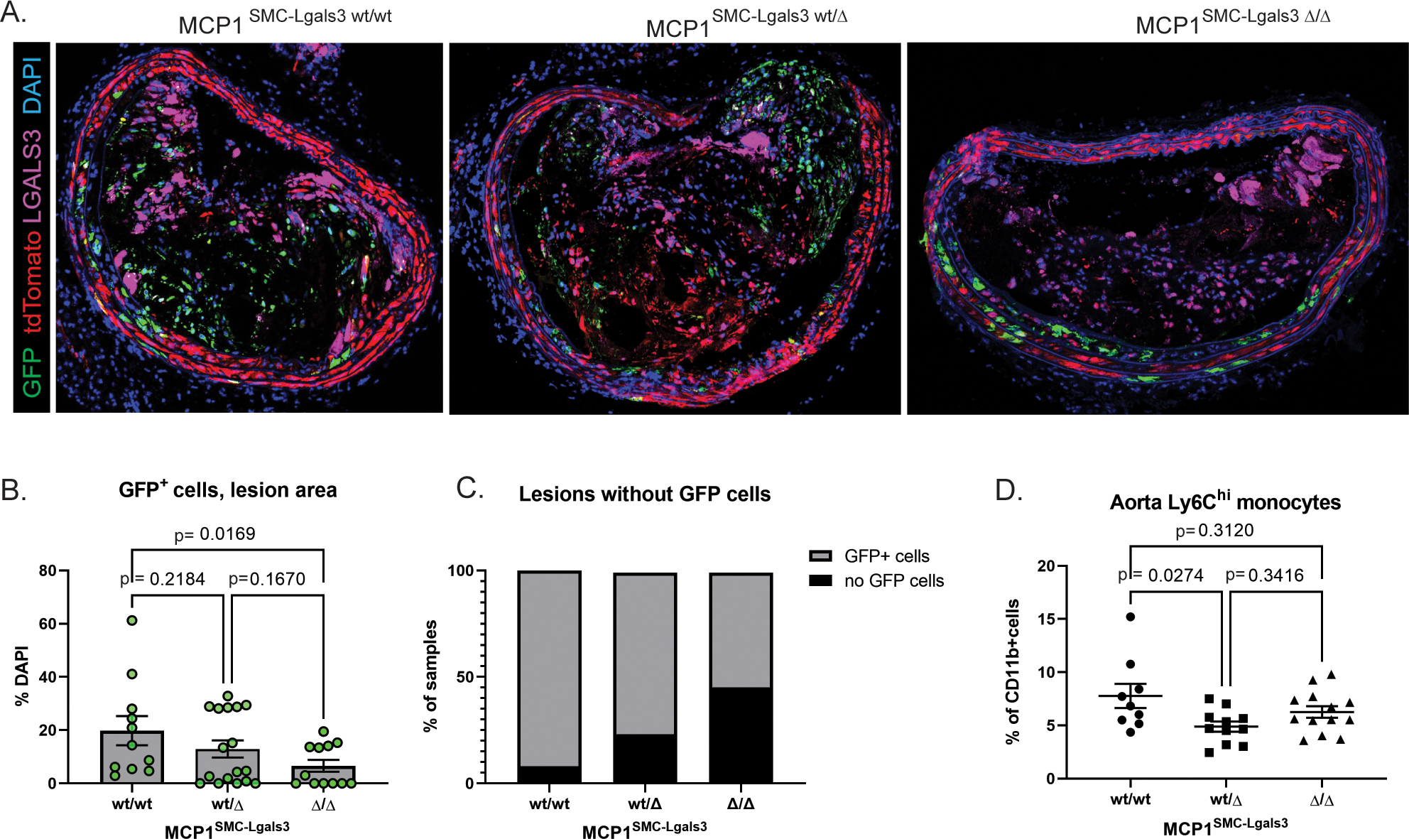
Figure 5.

MCP1 knockout in a subset of lesion SMC that transition through an Lgals3+ activation state results in decreased SMC phenotypic transition and aortic inflammation. A) BCA lesions from MCP1SMC-Lgals3 wt/wt, MCP1SMC-Lgals3 wt/Δ, and MCP1SMC-Lgals3 Δ/Δ mice fed a Western diet for 18 weeks were immunostained for GFP, tdTomato, LGALS3, and DAPI. B)Total GFP+ cells per DAPI were counted manually in 1μm sections from confocal images in Panel A. n=12–15, p-values refer to pairwise comparisons with Kruskal-Wallace test. C) BCA sections from Panels A-B were classified as a categorical variable based on entry or non-entry of GFP+ cells into lesions. Chi-squared test p=0.0001. D) Aortas from MCP1SMC-Lgals3 wt/wt, MCP1SMC-Lgals3 wt/Δ, and MCP1SMC-Lgals3 Δ/Δ mice fed a Western diet for 18 weeks were digested, stained for lymphocyte markers, and analyzed by flow cytometry. Ly6Chi monocytes were gated out of live, single CD45+CD11b+LY6G− leukocytes using fluorescence-minus-one controls. Despite being unchanged in peripheral blood at 18 weeks of Western diet, Ly6Chi monocytes were decreased in aortas of MCP1SMC-Lgals3 wt/Δ mice. n=10–12, p-values refer to Sidak multiple comparison test after one way ANOVA.
