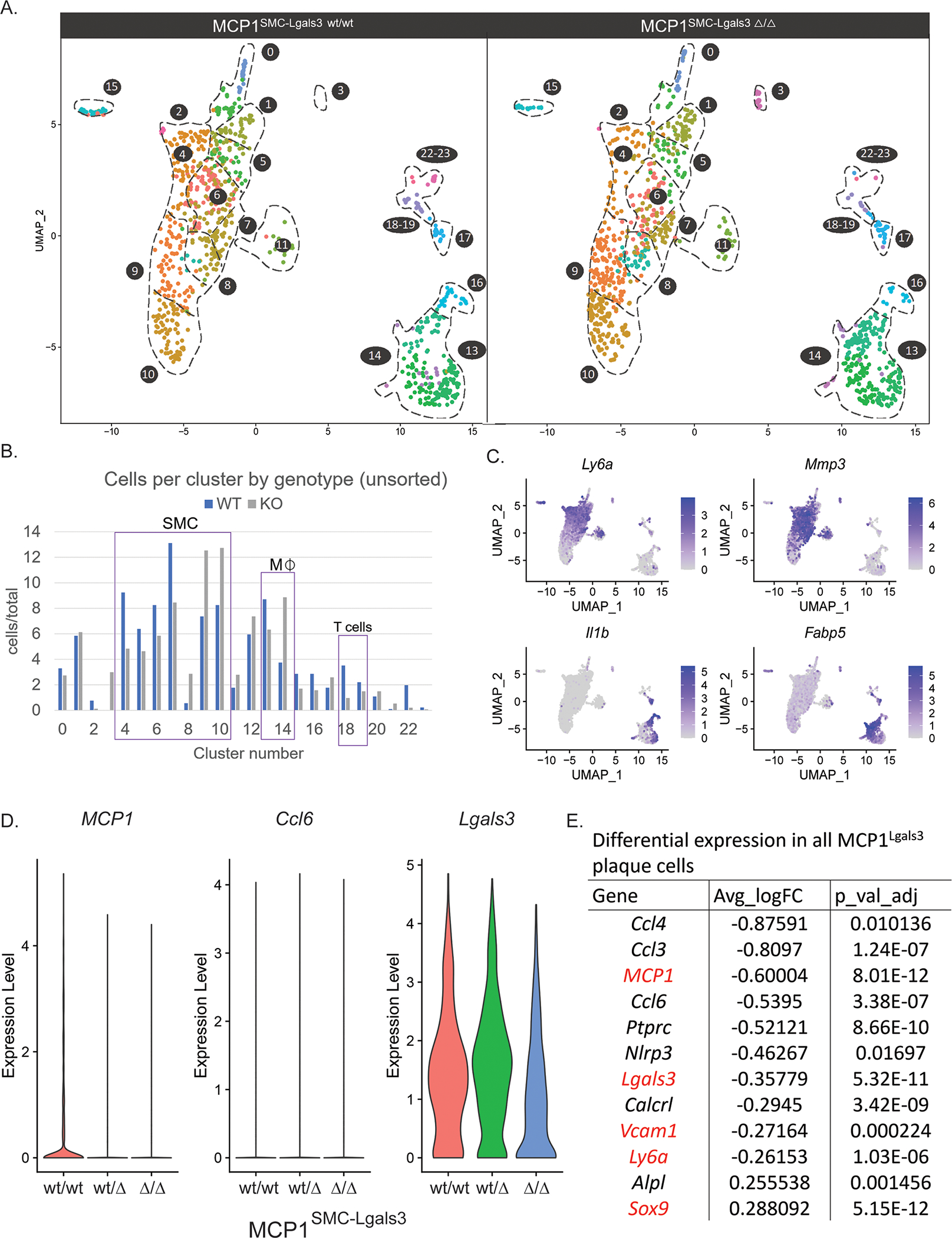
Figure 6.

MCP1 knockout in Lgals3-transitioned SMC resulted in decreased frequency of stem-like SMC and changes to multiple immune cell clusters A) Atherosclerotic lesions from MCP1SMC-Lgals3 wt/wt, MCP1SMC-Lgals3 wt/Δ, and MCP1SMC-Lgals3 Δ/Δ mice fed a Western diet for 18 weeks were microdissected from the BCA and single-cell RNAseq was performed on unsorted lesion samples, as well as GFP+ and tdTomato+ cells sorted from those lesions. 4 animals per genotype were pooled to make 9 total libraries representing 12 animals. 5,111 total cells passed quality control for this experiment and were clustered together, with cells from MCP1SMC-Lgals3 wt/wt and MCP1SMC-Lgals3 Δ/Δ mice shown in UMAP graphs in A. B) Cluster frequency between MCP1SMC-Lgals3 wt/wt and MCP1SMC-Lgals3 Δ/Δ mice was compared by measuring total cells in each cluster normalized for total cells in each genotype, using unsorted lesion samples to avoid enrichment bias. C) Feature plot diagrams showing clusters with expression of stem-like (Ly6a, clusters 4–5), ECM-remodeling (Mmp3, cluster 6–7), and macrophage (Il1b, clusters 13 and 16, Fabp5, cluster 14) genes. D-E) Single cell data from from experiments in A was normalized and differential expression analysis was done using MAST. D shows violin plots of normalized expression levels of three inflammatory genes in plaque cells from MCP1SMC-Lgals3 wt/wt, MCP1SMC-Lgals3 wt/Δ, and MCP1SMC-Lgals3 Δ/Δ mice. E shows an abbreviated chart of significantly differentially expressed genes between MCP1SMC-Lgals3 wt/wt and MCP1SMC-Lgals3 Δ/Δ plaque cells.
