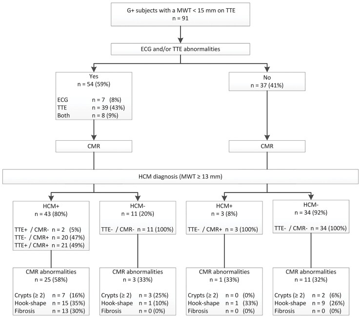
Figure 3.
Yield of CMR imaging following ECG and TTE. ECGs were abnormal in case of positive Cornell criteria or Romhilt–Estes score ≥4, TTEs were abnormal in case of LVH (MWT >10 mm) or LVOT obstruction. CMR reclassifies 8% of those without ECG/TTE abnormalities. CMR reveals additional abnormalities in all groups, but mostly in those with HCM.

