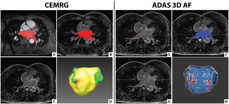
Figure 1.
Segmentation of the LA using CEMRG and ADAS 3D LA. (A–D) Screenshots taken in CEMRG. (A) LA segmentation (in red) on the 3D CE-MRA image using a thresholding tool. (B) Segmented LA (in red) co-registered with the 3D LGE image. (C) LA wall contour (in white) projected on the 3D LGE image. (D) 3D model of the LA incorporating fibrosis (red) after exclusion of the pulmonary veins and LA appendage. (E–H) Screenshots taken in ADAS 3D LA. (E) LA segmentation (in red) on the 3D LGE image by contouring the mid-LA wall. (F) LA segmentation (in blue) projected on the 3D LGE image. (G) LA wall contour (in blue) projected on the 3D LGE image. (H) 3D model of the LA incorporating fibrosis (red) after exclusion of the pulmonary veins and LA appendage.

