. 2022 Feb 17;28(8):1572–1585. doi: 10.1158/1078-0432.CCR-21-3324

Figure 1.

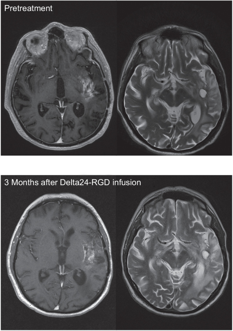
Figure 1. Illustrative case patient 7, a 67-year-old male with a second recurrence of a GBM, diagnosed 13 months earlier. Virus infusion of 1 × 10⁹ vp was uncomplicated, but patient was readmitted 3 months later due to neurological deterioration. MR showed a decrease of the contrast-enhancing part of the tumor but an increase of the signal intensity in the peritumoral area on the T2- and FLAIR-images, interpreted as increased edema and stable tumor (left side: T1 after gadolinium; right side: T2 images). During hospitalization, patient deteriorated rapidly, dying with clinical signs of sepsis. At autopsy a diverticulitis with sigmoid perforation was found. Brain autopsy revealed a relatively small tumor in relation to the MR images, containing immune cell infiltrates (Fig. 5A), necrotic areas, infarctions, and absence of vascular proliferation.

Illustrative case patient 7, a 67-year-old male with a second recurrence of a GBM, diagnosed 13 months earlier. Virus infusion of 1 × 10⁹ vp was uncomplicated, but patient was readmitted 3 months later due to neurological deterioration. MR showed a decrease of the contrast-enhancing part of the tumor but an increase of the signal intensity in the peritumoral area on the T2- and FLAIR-images, interpreted as increased edema and stable tumor (left side: T1 after gadolinium; right side: T2 images). During hospitalization, patient deteriorated rapidly, dying with clinical signs of sepsis. At autopsy a diverticulitis with sigmoid perforation was found. Brain autopsy revealed a relatively small tumor in relation to the MR images, containing immune cell infiltrates (Fig. 5A), necrotic areas, infarctions, and absence of vascular proliferation.