. 2022 Feb 17;28(8):1572–1585. doi: 10.1158/1078-0432.CCR-21-3324

Figure 5.

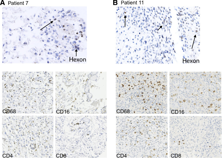
Figure 5. IHC analysis of post-mortem tumor material of patient 7 (A, 3 months after virus administration, left) and re-resection material of patient 11 (B, 4 weeks after virus administration, right) for the adenoviral protein hexon (top images), showing scattered hexon-positive cells and for the presence of immune cells (bottom images); CD68+ cells (macrophage marker), CD16+ (monocyte marker), and CD4+ lymphocytes are quite abundant in both samples. CD8+ lymphocytes are less frequent in both samples.

IHC analysis of post-mortem tumor material of patient 7 (A, 3 months after virus administration, left) and re-resection material of patient 11 (B, 4 weeks after virus administration, right) for the adenoviral protein hexon (top images), showing scattered hexon-positive cells and for the presence of immune cells (bottom images); CD68+ cells (macrophage marker), CD16+ (monocyte marker), and CD4+ lymphocytes are quite abundant in both samples. CD8+ lymphocytes are less frequent in both samples.