. 2022 Feb 17;28(8):1572–1585. doi: 10.1158/1078-0432.CCR-21-3324

Figure 7.

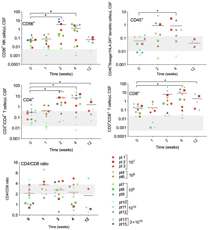
Figure 7. Immune cells were isolated from cerebrospinal fluid at indicated timepoints after Delta24-RGD infusion and analyzed by flow cytometry with the following markers: CD45+CD3−CD56+ (NK cells), DCs (CD45+/lineage−/HLA-DR+), CD45+CD3+/CD4+ (CD4+ T-lymphocytes), and CD45+CD3+/CD8+ (CD8+ T-lymphocytes). Immune cell subsets are presented as absolute numbers per μL CSF. Gray-shaded areas indicate the normal range for each cell type.

Immune cells were isolated from cerebrospinal fluid at indicated timepoints after Delta24-RGD infusion and analyzed by flow cytometry with the following markers: CD45+CD3CD56+ (NK cells), DCs (CD45+/lineage/HLA-DR+), CD45+CD3+/CD4+ (CD4+ T-lymphocytes), and CD45+CD3+/CD8+ (CD8+ T-lymphocytes). Immune cell subsets are presented as absolute numbers per μL CSF. Gray-shaded areas indicate the normal range for each cell type.