
Figure 2.
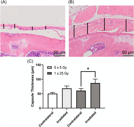
Irradiated capsule was significantly thicker in the mice that received a single dose of 25 Gy. Hematoxylin and eosin (H&E)‐stained slides of coronal sections show healthy joint capsule from the contralateral leg of a mouse that received fractionated doses of radiation (A) and inflamed capsule tissue from the irradiated leg of a mouse that received a single bolus dose of radiation (B). Capsular thickness is indicated by vertical black bars, scale bar is 50 µm. Representative images were taken from the proximal edge of the femur (lower half of image) with the capsular and synovial tissue in the center of the image. Images were analyzed using ImageJ length measurement tool to determine average capsule thickness in µm (C). Dotted bars indicate irradiated legs, clear bars indicate contralateral legs. 5 × 5 Gy (n = 8); 1 × 25 Gy (n = 5); *p < 0.05, two‐way analysis of variance (ANOVA) [Color figure can be viewed at wileyonlinelibrary.com]