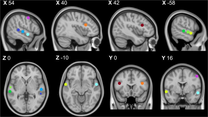
FIGURE 1.
Temporal voice area fMRI localizer task results: Purple = right premotor cortex, dark blue = right posterior temporal gyrus, middle blue = right middle temporal gyrus, light blue = right anterior temporal gyrus, orange = right inferior frontal cortex, dark green = left posterior superior temporal gyrus, middle green = left middle superior temporal gyrus, light green = left anterior superior temporal gyrus, and red = left inferior temporal cortex. All coordinates listed in MNI space (x,y,z). This image was created using the FSL toolbox fsleyes (McCarthy, 2022).

