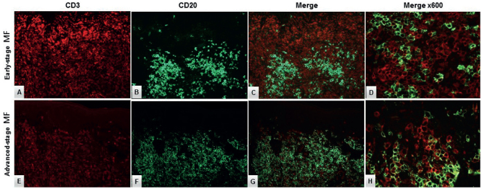
Fig. 4.
Double-immunofluorescence staining with CD3 (red) and CD20 (green) in (A–D) early-stage (ES) mycosis fungoides (MF) and (E–H) advanced-stage (AS) MF. The neoplastic T-cells did not display aberrant CD20 expression (expected yellow reaction product) in (D) ES-MF or (H) AS-MF, respectively. Original magnification ×200 in A–C, E–G and ×600 in D, H.

