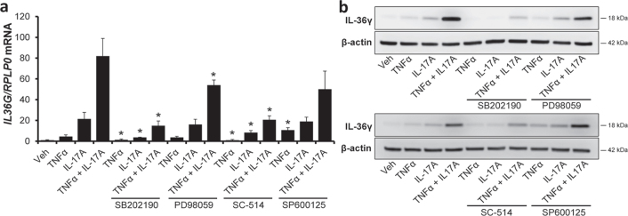
Fig. 2.
Characterization of tumour necrosis factor-α (TNFα)/interleukin (IL)-17A-induced IL-36γ expression. Human keratinocytes were incubated with the p38 mitogen-activated protein kinase (MAPK) inhibitor SB202190, the ERK1/2 inhibitor PD98059, the IKK2 inhibitor SC-514, the JNK inhibitor SP600125 or with vehicle (Veh) for 45 min before stimulation with TNFα and/or IL-17A for 24 h. (a) IL36G mRNA expression was measured by qPCR (n = 3). Results are expressed as mean ± standard deviation. RPLP0 mRNA expression was used for normalization. (b) The protein level of IL-36γ was measured by western blotting with β-actin as a loading control (n = 3). *p < 0.05 compared with TNFα- and/or IL-17A-stimulated cells without inhibitors.

