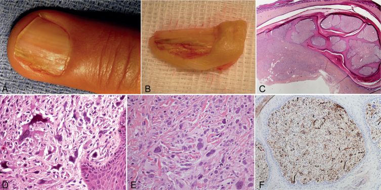
Fig. 1.
(A) Fibrokeratoma-like features with longitudinal xanthonychia. (B) Macroscopic examination showing the multibranched fibrokeratoma feature. (C) Scanning power of the transverse section of the matrical region, under the dorsal portion of the proximal nail fold, showing the polypoid feature with multiple fibroepithelial digitations. Note the infiltrative growth of the tumour (asterisk), and partial epidermal metaplasia of the matrix epithelium at the level of the tumour. The medial and peripheral part of the lesion with a thin nail plate is indicated by an arrow (original magnification (OM) ×20). (D) Higher magnification of the enlarged hyperchromatic bizarre-appearing nuclei (OM ×400). (E) Xanthomatous cells interspersed between cells with granular cytoplasm (OM ×400). (F) Diffuse CD34 expression of both the spindle and epithelioid cells (OM ×100).

