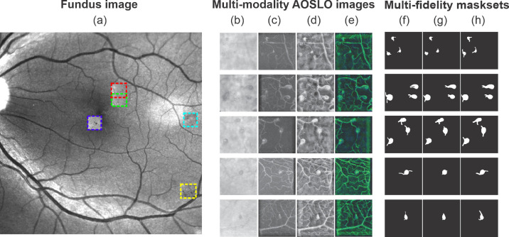
Figure 1.
Multi-modality AOSLO images and multifidelity mask sets are used to train and test the AOSLO-net. (a) Five MAs imaged by AOSLO (highlighted by boxes in different colors) superimposed on a digital fundus photograph from an eye with diabetic retinopathy. (b–e) Examples of four sets of AOSLO images with different modalities used to train and test the AOSLO-net: (b) raw images extracted from the AOSLO videos; (c) perfusion maps (also see Supplementary Fig. S1); (d) preprocessed AOSLO images with enhancement; (e) two-modality images which are generated by concatenating the perfusion maps (c) and enhanced AOSLO images (d). Details of how multimodality AOSLO images are generated can be found in the Method section. These images illustrate that a varied number of MAs can be detected in a single AOSLO image. Images in row 1–3 contain multiple MAs, whereas images in row 4-5 contain a single MA with complicated background vessels whose size may be comparable to the MA. (f–h) Three sets of masks are generated independently to examine the robustness of the AOSLO-net to mask sets with different qualities. Normal set (f): masks are created based on both the AOSLO videos and perfusion maps to illustrate the body of MAs and their feeding and draining vessels. Short set (g): masks are designed to show shorter feeding and draining vessels of MAs compared to the normal set, while the thickness of the vessels remains similar to the normal masks. Thick set (h): masks are designed to show thicker feeding and draining vessels of MAs compared to the normal set, while the length of the vessels remains similar to the normal masks.

