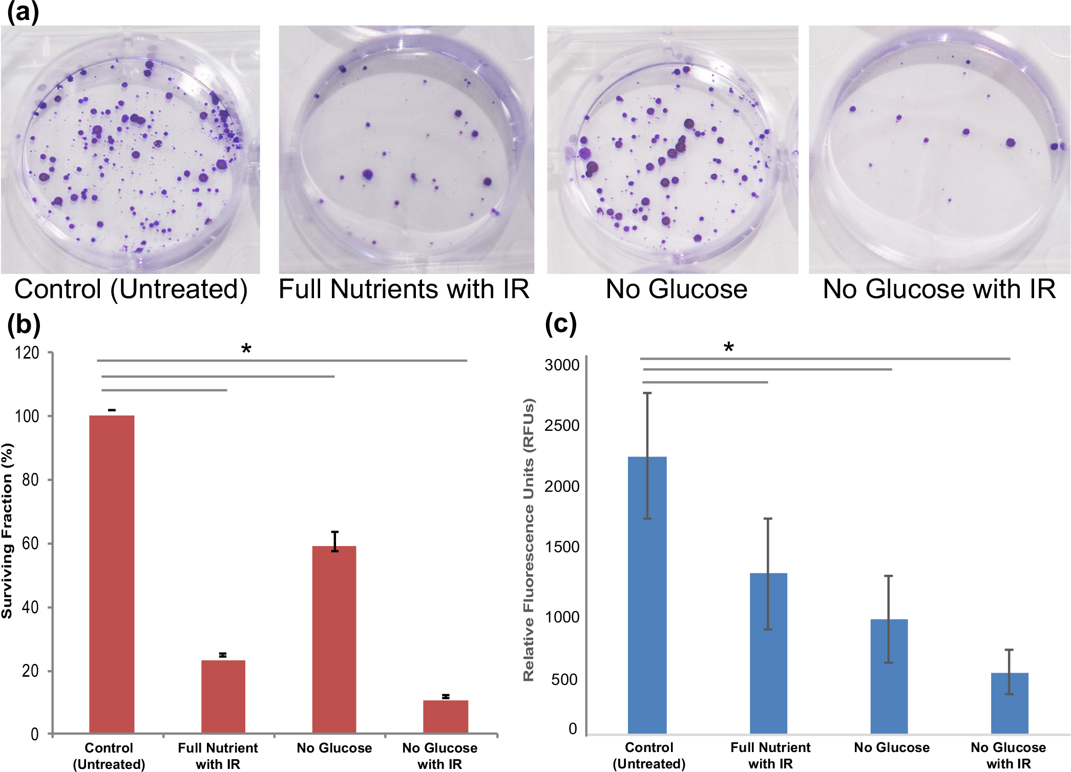
Figure 3.

Phenotypic assays to assess clonogenicity and cell viability of treatment on HCT (a) Images of clonogenicity assay in a six-well plate after fiv e doublings of HCT 116 cells. (b) Surviving fraction (%) from clonogenicity assay compared to the full nutrient control, to determine ability of cells to proliferate after treatment. (c) Cell viability assay results reporting cell viability of spheroids after treatment in relative fluorescence units (RFUs) using the Promega cell titer blue assay
