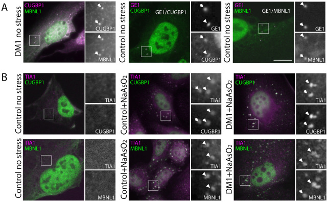
Fig. 1.
MBNL1 and CUGBP1 both localise to P-bodies within HLECs and to SGs following treatment with NaAsO2. (A) Endogenous MBNL1 (left panel, green on overlay) and CUGBP1 (left panel, magenta on overlay) colocalise in punctate cytoplasmic structures (arrows) despite showing little colocalisation in the nucleus (see also Fig. S1). Counterstaining with antibodies against the P-body marker GE1 (centre and right panels, magenta on overlay) demonstrates that both CUGBP1 (centre panel, green on overlay) and MBNL1 (right panel, green on overlay) localise to P-bodies (arrows) in DM1 and control cell lines (see also Fig. S1). (B) SGs detected with antibodies against TIA1 (magenta on overlay) are not seen in unstressed cells (left panels). Following treatment with 0.5 mM NaAsO2 for 45 min, control cells (centre panels) and DM1cells (right panels) show clear cytoplasmic SGs (arrows) identified by antibodies against TIA1 (magenta on overlays) containing both CUGBP1 (top row green on overlays) and MBNL1 (bottom row, green on overlays; see also Fig. S1). Boxed areas are shown magnified at the right of each panel; scale bar: 7 µm.

