Fig. 8.
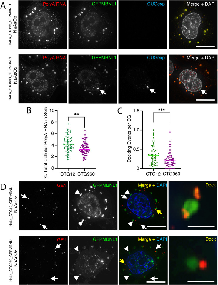
Reduced amounts of poly(A) RNA in SGs, and altered docking events between P-bodies and SGs suggests defects in their function in cells expressing CUGexp RNA (A) RNA FISH of HeLa_CTG12_GFPMBNL1 (top) and HeLa_CTG960_GFPMBNL1 (bottom) cells treated for 90 min with NaAsO2. Poly(A) RNA (first images; red in merged images) accumulates in the nucleus and in cytoplasmic SGs of both cell lines. CUGexp RNA (third images; blue in merged images) is detected only in the nuclear foci (arrows) of HeLa_CTG960_GFPMBNL1 cells. GFP-tagged MBNL1 (GFPMBNL1) accumulates in SGs of both cell lines and in the CUGexp foci when they are present (second images; green in merged images). (B) The total amount of cellular poly(A) RNA (in %) in SGs is reduced in cells that express CUGexp RNA (CTG960) than in control cells (CTG12). Data shown are pooled from two independent experiments. n=71 (CTG12), n=84 (CTG960); **P<0.01, unpaired t-test. (C) The number of docking events between SGs and P-bodies is lower in cells that express CUGexp RNA (CTG960) than in control cells (CTG12). Data shown are pooled from two independent experiments (CTG12, n=57 cells; CTG960, n=60 cells); ***P<0.001, Mann–Whitney U test. (D) HeLa_CTG12_GFPMBNL1 (top) and HeLa_CTG960_GFPMBNL1 (bottom) cells, treated with NaAsO2 for 90 min. P-bodies detected with antibodies against GE1 (indicated by white arrows in first and third images; shown in red in merged and magnified images), show docking events (indicated by yellow arrows in merged images and shown magnified in panels on the right). SGs are detected by staining for GFPMBNL1 (indicated by arrowheads and shown in green in merged images). All nuclei are counterstained with DAPI (white in merged image in A; blue in merged image in B). Scale bars: 10 µm (A,D); 2 µm (magnified images in D).

