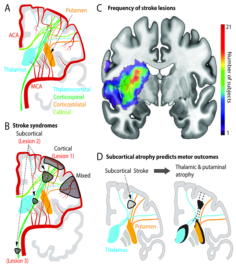
Figure 1. Vascular and structural anatomy of stroke syndromes.

A. Illustration of the anterior vascular circulation, cortical and deep grey nuclei and connecting fibers. ACA=anterior cerebral artery; MCA=middle cerebral artery.
B. Common symptomatic stroke lesion types. Lesion 1 is pure cortical. Lesion 2 is in corona radiata (“subcortical”). Lesion 3 is the brainstem. A mixed cortical and subcortical lesion is also marked. These lesions are also referred to in Figure 2.
C. Human brain MRI with superimposed stroke “lesion density” map. Lesions previously published in (Lin et al., 2019) and (Lin et al., 2021). Colorbar illustrates the number of subjects with a lesion in that region in a cohort of n=65 patients with upper extremity deficits.
D. Atrophy in subcortical nuclei (thalamus, putamen) can emerge due to disconnection of these nuclei from cortex. Atrophy is associated with persistent motor deficits.
