
Figure 3: Motor maps and motor evoked potential.
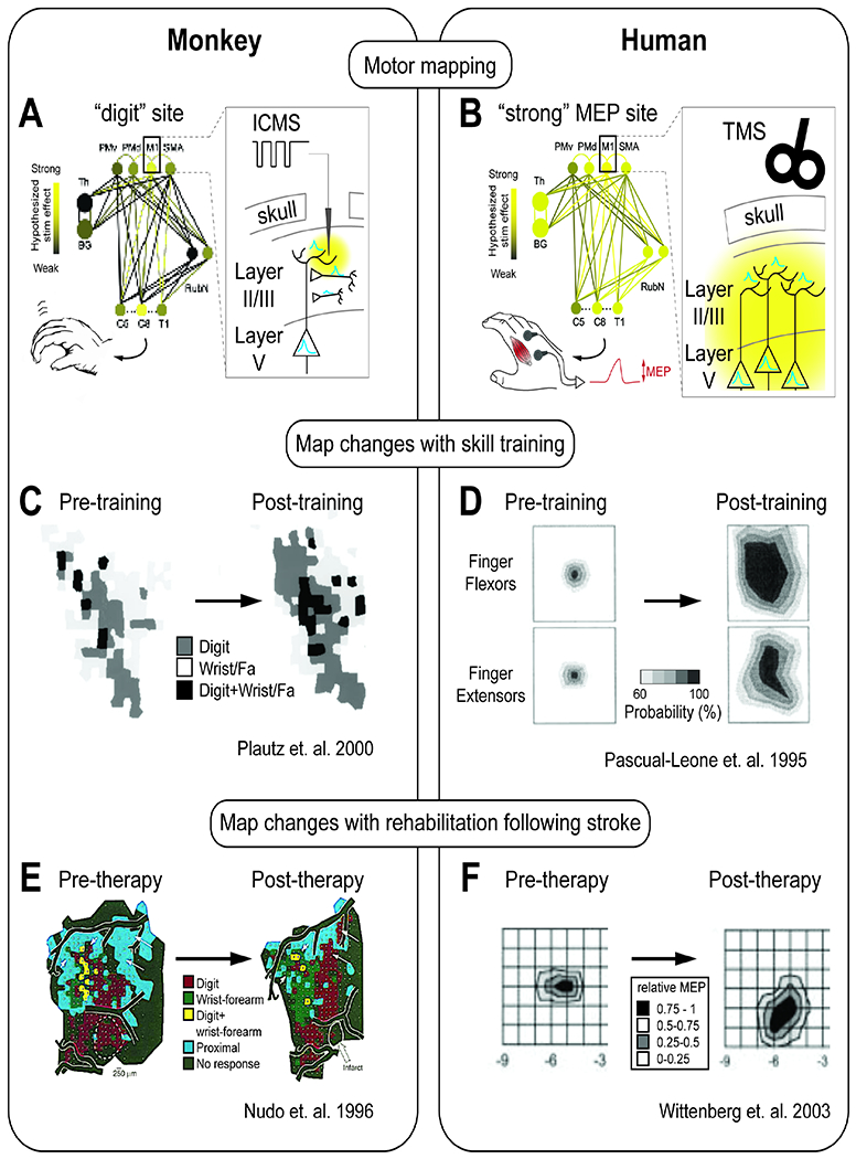
A. Intracortical microstimulation (ICMS) derived motor maps performed in preclinical studies. Microelectrodes are introduced into different cortical sites. Stimulation pulses are delivered, and observations of which limbs move are noted.
B. Transcranial magnetic stimulation (TMS) derived motor maps performed in humans. TMS coils are positioned to activate specific cortical sites. Stimulation is delivered, and muscle activity at one site is monitored to capture “motor-evoked potentials” (MEPs).
C. Training of a skilled RTG movement increases the digit, wrist, and forearm representation in motor maps (Plautz 2000).
D. Training of a skilled hand movement over 5 days increases the probability of evoking a motor potential (left = day 1, right = day 5) (Pascual-Leone 1995).
E. Following rehabilitation therapy after an infarct, an expansion of the evoked digit representation in the cortical map (Nudo, 1996)
F. Following rehabilitation therapy, an expansion of areas of cortex that can evoke an MEP (Wittenberg, 2003)