
Figure 8. Evolution of resting, task and NREM sleep activity after stroke.
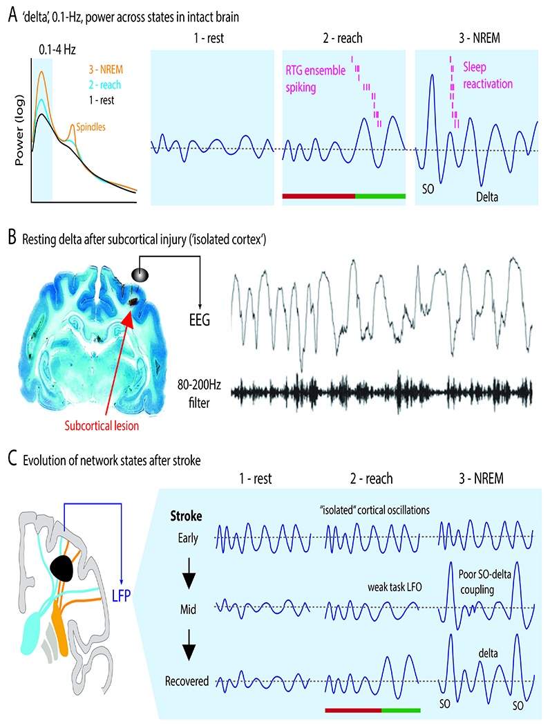
A. Activity patterns in the delta band across three network states. There is growing evidence that spiking activity linked to task LFOs is also reactivated during delta band slow-wave activity during sleep. Arrowhead indicates movement onset. Color for preparation vs execution as prior.
B. Reactivations in NREM sleep are prominent during slow oscillations (SO). Reactivations are detected using templates created – using dimensionality reduction methods, i.e., latent factors or LFs – from task related activity.
C. Increased resting state delta after a subcortical lesion that isolates cortex. This can be seen in both hemispheres.
D. Model of changes in delta patterns with recovery. Early states are associated with ‘autonomous’ delta waves that largely do not vary across states, including sleep. With late recovery, it possible for normal cortico-thalamic interactions and normal transitions between states. SO=slow oscillation.