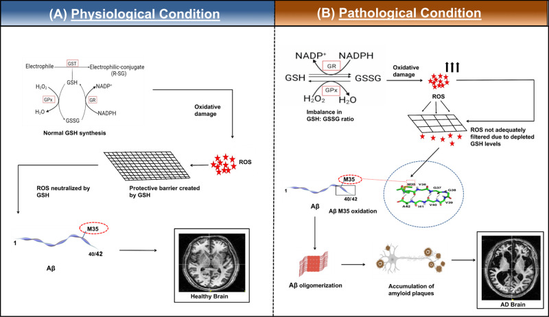
Figure 2.
Schematic representation of the impact of GSH in (A) healthy brain and (B) pathological condition in AD brain. (A) In a normal brain under physiological conditions, glutathione levels are maintained. GSH participates in the GSH cycle.89 The GSH:GSSG ratio is maintained at 100:1 via the cycle by recycling GSH from oxidized glutathione (GSSG) intracellularly. ROS is actively detoxified by GSH, which acts as a modulator for the radicals, and ROS cannot cause further oxidation of the M35 residue of Aβ. The redox homeostasis is thus maintained in the healthy brain. (B) A model of Alzheimer’s disease pathology due to oxidative stress. Depleted GSH levels can lead to increased levels of free radicals. These free radicals can then oxidize the M35 residue leading to several biochemical modifications12 like lipid peroxidation and protein modifications as well as formation of amyloid plaques, which can consequently form neurofibrillary tangles. Red star indicates ROS and oxidative damage. Representative image of structure of residues 35–42 of Aβ1–42 has been adapted with permission from ref (79). Copyright 2008 Elsevier B.V. Representative MRI images of healthy and AD brain are taken from NINS laboratory data.

