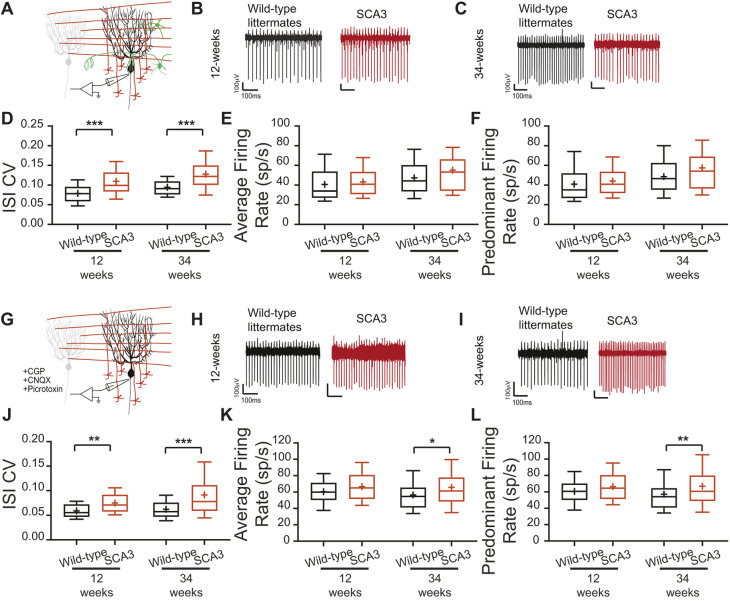
Fig. 4.
Synaptic and intrinsic components to Purkinje cell irregularity. (A-L) In vitro recordings were performed at 12 and 34 weeks of age, with synaptic transmission intact (A-F) and synaptic transmission blocked (G-L). Example Purkinje cell firing traces from wild-type (black) and SCA3 (red) mice at 12 weeks (B,H) and 34 weeks (C,I) of age. When synaptic transmission is intact (A-F), Purkinje cell ISI CV (C) is increased in SCA3 mice at both time points, with no change in average (E) and predominant (F) firing rate. When synaptic transmission is blocked (G-L), Purkinje cell ISI CV (J) is increased in SCA3 mice at both time points, with no change in average (K) and predominant (L) firing rate at 12 weeks, but an increase in both at 34 weeks. Synaptic transmission intact: 12 weeks: wild-type N=4, n=38; SCA3 N=4, n=50. 34 weeks: wild-type N=16, n=89; SCA3 N=17, n=91. Synaptic transmission blocked: 12 weeks: wild-type N=3, n=22; SCA3 N=3, n=29. 34 weeks: wild-type N=13, n=92; SCA3 N=15, n=121. *P<0.05, **P<0.01, ***P<0.001. Unpaired, two-tailed Student's t-test (K, 12 weeks; L, 12 weeks), Mann–Whitney test (D, all; E, all; J, all; K, 34 weeks; L, 34 weeks).

