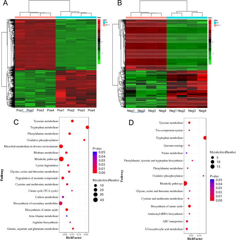
Fig. 4.
Classification and analysis of differential metabolites in P. polymyxa SC2-M1. Cluster analysis of differential metabolites in the positive (A) and negative (B) ion mode. Each row represents a differential metabolite and each column represents a sample. The color represents the expression level of differential metabolites, and the green to red corresponds to the expression level from low to high. Bubble chart of metabolic pathway enrichment analysis in the positive (C) and negative (D) ion mode. Red represents the significant enrichment and the size of the dot represents the number of different metabolites annotated in the pathway

