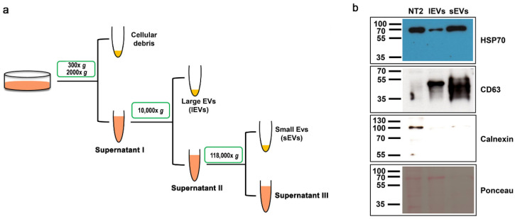
Figure 1.
Isolation of extracellular vesicles from NTERA2 cells. (a) General outline of the differential centrifugation-based protocol used. (b) Protein immunoblotting with the antibody against the EV specific markers heat shock protein 70 (HSP70) and CD63, and the negative EV marker calnexin. Equal protein amounts (20 ug) of NTERA2 cell lysates (NT2), large extracellular vesicle (lEV), and small extracellular vesicle (sEV) preparations were analyzed. On the bottom: the filter after protein transfer and Red Ponceau protein revelation.

