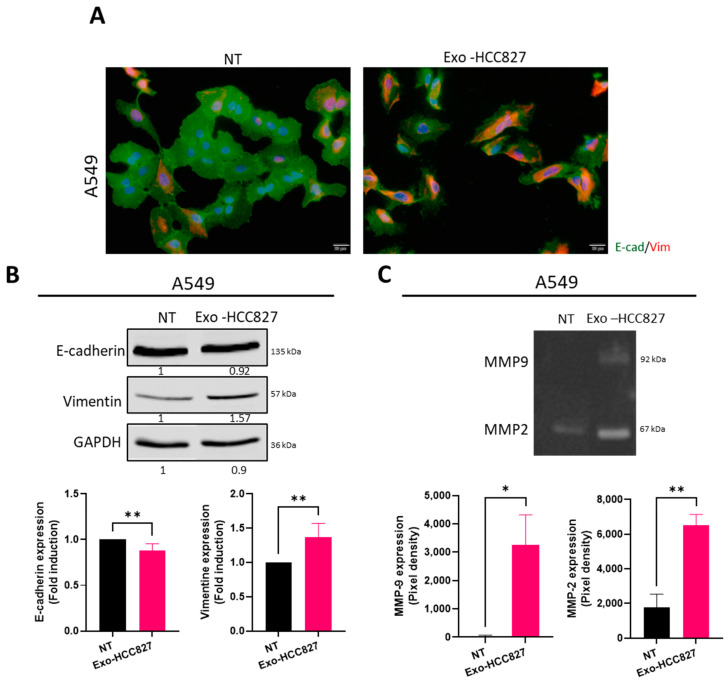
Figure 4.
Exosomes derived from the HCC827 cell line promoted extracellular matrix degradation and EMT (A) Immunofluorescence detection of vimentin (red) and E-cadherin (green) in A549 cells treated with exosomes isolated from HCC827 supernatant. Nuclei were stained with DAPI (blue). Scale bar = 20 μm. (B) Western blot analysis and quantification of vimentin and E-cadherin expression in A549 cells treated with exosomes isolated from HCC827 supernatant. GAPDH was used as a loading control (mean ± SEM, n = 4; statistical analysis was performed using unpaired t-test). (Original whole Western-blot Images and densitometry reading in Figure 3 and Table S4). (C) Zymography analysis and quantification of A549 cells (right) treated with exosomes isolated from HCC827 supernatant (mean ± SEM, n = 4; statistical analysis was performed using unpaired t-test). Significance was set at * p < 0.05 and ** p < 0.01.

