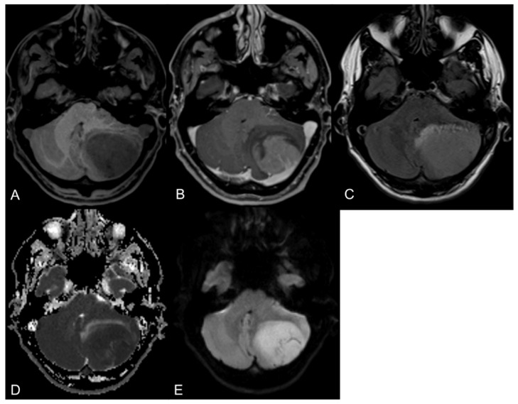
Figure 1.
Axial MR images of a 46-year-old male patient with a medulloblastoma located in the left cerebellar hemisphere. The tumor shows a characteristic hypointense signal on T1-weighted images (A) with a mainly homogenous contrast enhancement (B), a hyperintense signal on FLAIR images (C) and a considerable diffusion restriction (D,E).

